CPT Code 47000⁚ Liver Biopsy
CPT code 47000 signifies a percutaneous needle biopsy of the liver, a procedure extracting a liver tissue sample for diagnosis․ This can be performed with various imaging guidance, such as CT or ultrasound, to ensure accurate needle placement and sample acquisition․ The procedure is commonly used to assess liver disease and lesions․
Percutaneous Liver Biopsy Procedures
Percutaneous liver biopsy, a minimally invasive procedure, involves inserting a thin needle through the skin to obtain a liver tissue sample․ This technique offers several advantages over open surgical biopsy, including reduced invasiveness, shorter recovery times, and lower risk of complications․ The procedure is guided by imaging techniques, most commonly ultrasound or CT scans․ Ultrasound guidance is frequently preferred for its real-time visualization and safety profile․ However, CT guidance may be necessary for visualizing lesions not easily seen with ultrasound, particularly deep-seated lesions or those obscured by overlying structures․ The choice of guidance modality depends on the specific clinical scenario, lesion characteristics, and patient factors․
Before the procedure, the patient undergoes a thorough evaluation, including assessment of coagulation parameters to minimize bleeding risk․ Local anesthesia is administered to numb the biopsy site, and the procedure is typically performed under close monitoring․ After the biopsy, the patient is observed for potential complications such as bleeding or pain․ The obtained tissue sample is then sent to a pathology laboratory for histopathological analysis, providing crucial information for diagnosis and treatment planning․ Post-procedure care includes monitoring vital signs and managing any discomfort․ Overall, percutaneous liver biopsy is a valuable tool in the diagnostic workup of various liver conditions․
CT Guidance and CPT Codes
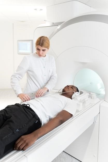
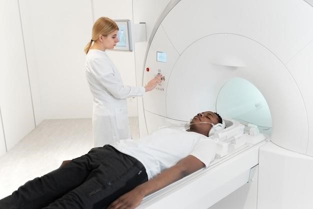
Computed tomography (CT) guidance plays a crucial role in percutaneous liver biopsies, particularly when targeting specific lesions or when ultrasound visualization is insufficient․ CT scans provide detailed cross-sectional images of the liver, allowing precise localization of the target area and accurate needle placement․ This is especially beneficial for deep-seated lesions, those obscured by gas or other structures, or when a higher degree of accuracy is required․ The integration of CT guidance enhances the safety and efficacy of the procedure by minimizing the risk of complications such as bleeding or pneumothorax․
The use of CT guidance is reflected in the billing and coding associated with the procedure․ While CPT code 47000 represents the liver biopsy itself, additional codes may be necessary to capture the imaging guidance component․ Specific codes depend on the imaging services performed․ For instance, code 77012 is often used to represent CT guidance for needle placement․ Accurate coding is crucial for appropriate reimbursement and reflects the complexity and precision involved in CT-guided liver biopsies․ Understanding the specific CPT codes for CT guidance and their appropriate application is vital for accurate medical billing and record-keeping․
Additional CPT Codes for Imaging Guidance
Beyond CT guidance (often coded as 77012), other imaging modalities can guide liver biopsies, necessitating additional CPT codes for accurate billing․ Ultrasound, a common alternative, uses high-frequency sound waves to visualize the liver, guiding needle placement․ The appropriate CPT code for ultrasound guidance is 76942․ Magnetic resonance imaging (MRI), offering superior soft tissue contrast, may be used in specific cases, although less frequently than CT or ultrasound․ The CPT code for MRI guidance is 77021․ The selection of the appropriate code depends entirely on the imaging modality employed․ Incorrect coding can lead to claim denials or underpayment․
It’s crucial to note that these imaging guidance codes are typically reported in addition to the primary biopsy code (47000)․ Bundling rules may apply, depending on payer policies and the specific circumstances of the procedure․ Consult the most current CPT codebook and payer guidelines to ensure accurate and compliant billing practices․ Proper documentation of the imaging modality used is critical for supporting the billed codes․ Always verify payer-specific requirements before submitting claims, as coding practices may vary․ Clear and precise documentation ensures proper reimbursement for the services rendered․

Types of Liver Biopsies
Liver biopsies are categorized as either non-focal (assessing general liver health) or focal (targeting specific lesions)․ The choice influences the imaging guidance (ultrasound or CT) and procedural approach, affecting both the technique and the CPT codes used for billing․
Non-Focal vs․ Focal Biopsies
The distinction between non-focal and focal liver biopsies significantly impacts the procedure and associated CPT codes․ Non-focal biopsies, also known as non-targeted biopsies, are performed to assess the overall health and condition of the liver parenchyma․ These are often used in the evaluation and staging of diffuse liver diseases such as non-alcoholic steatohepatitis (NASH), where a representative sample of the liver tissue is needed to determine the extent and severity of the disease․ In contrast, focal biopsies, or targeted biopsies, are directed at a specific area within the liver, such as a suspicious lesion or mass identified on imaging studies․ The goal of a focal biopsy is to obtain a tissue sample from this specific area for diagnostic purposes, such as determining whether a lesion is benign or malignant․ The choice between these two types of biopsies is made based on clinical findings and imaging results․ The location and nature of the target influence the selection of imaging guidance, whether ultrasound or CT, and the specific CPT codes utilized to report the procedure․
Ultrasound vs․ CT Guidance
The selection between ultrasound and computed tomography (CT) guidance for a liver biopsy depends largely on the clinical scenario and the nature of the lesion being targeted․ Ultrasound guidance is generally the preferred method for non-focal biopsies, aiming to obtain a representative sample of liver tissue․ Its advantages include its real-time imaging capabilities, portability, lower cost, and lack of ionizing radiation․ CT guidance, however, often proves superior when dealing with focal lesions, particularly those that are difficult to visualize or access using ultrasound․ CT provides excellent anatomical detail and allows for precise needle placement, especially in cases of deep-seated or small lesions․ In certain situations, a combined approach employing both ultrasound and CT may be employed․ For instance, if ultrasound initially identifies a lesion, CT might then be used for more precise targeting during the biopsy․ The choice of imaging modality directly impacts the CPT codes used for billing, with separate codes reflecting the type of guidance utilized․ The decision regarding the most appropriate guidance method rests upon a careful consideration of the specific clinical context and the characteristics of the target lesion․

Risks and Complications
While generally safe, liver biopsies carry potential risks, including bleeding (hemorrhage), infection, and pain․ Rare but serious complications can occur, such as bile duct injury or pneumothorax․ Post-procedure monitoring is crucial to detect and manage any complications promptly․
Potential Complications of Liver Biopsy
Liver biopsy, even when guided by CT, carries inherent risks․ While generally a safe procedure, several potential complications can arise․ These complications range in severity from minor discomfort to life-threatening events․ One of the most common complications is pain, often felt in the right upper abdomen or radiating to the right shoulder․ This pain is usually manageable with analgesics but can be severe in some cases․ Another significant risk is bleeding, which can range from minor oozing to significant hemorrhage․ The severity of bleeding depends on various factors, including the patient’s coagulation status and the location of the biopsy․ In rare instances, major bleeding can lead to the need for blood transfusions or even emergency surgery․ Infection at the biopsy site is another possibility, although less frequent with proper sterile technique․ This infection can manifest as localized pain, redness, swelling, or pus formation and may require antibiotic treatment․ Less common but potentially serious complications include pneumothorax (collapsed lung), bile duct injury, and seeding of tumor cells along the needle tract (in cases of malignancy)․ The likelihood of these complications is influenced by factors such as the patient’s overall health, the experience of the physician performing the procedure, and the specific technique employed․ Careful patient selection and meticulous technique are essential to minimizing these risks․
Post-Procedure Monitoring and Recovery
Post-procedure monitoring is crucial after a CT-guided liver biopsy to detect and manage potential complications promptly․ The patient’s vital signs, including heart rate, blood pressure, and oxygen saturation, are closely monitored․ Pain levels are assessed and managed with analgesics as needed․ The biopsy site is regularly inspected for any signs of bleeding or infection․ The observation period typically lasts several hours, with the frequency of monitoring adjusted based on the patient’s condition and risk factors․ In the initial hour, vital signs and the biopsy site are checked every 15 minutes․ This frequency gradually decreases to every 30 minutes for the next two hours, and then to hourly checks for the remainder of the observation period․ Patients are instructed to report any new or worsening pain, shortness of breath, or other concerning symptoms․ Before discharge, the patient’s vital signs should be stable, with no evidence of bleeding or hemodynamic instability․ Patients are advised to rest for the remainder of the day and avoid strenuous activity․ Specific instructions regarding diet, activity level, and follow-up appointments are provided․ The length of the recovery period varies depending on individual factors, but most patients experience a relatively quick recovery with minimal discomfort; However, patients should be aware of the potential for delayed complications and instructed to contact their healthcare provider if any concerning symptoms develop․


